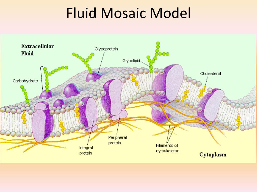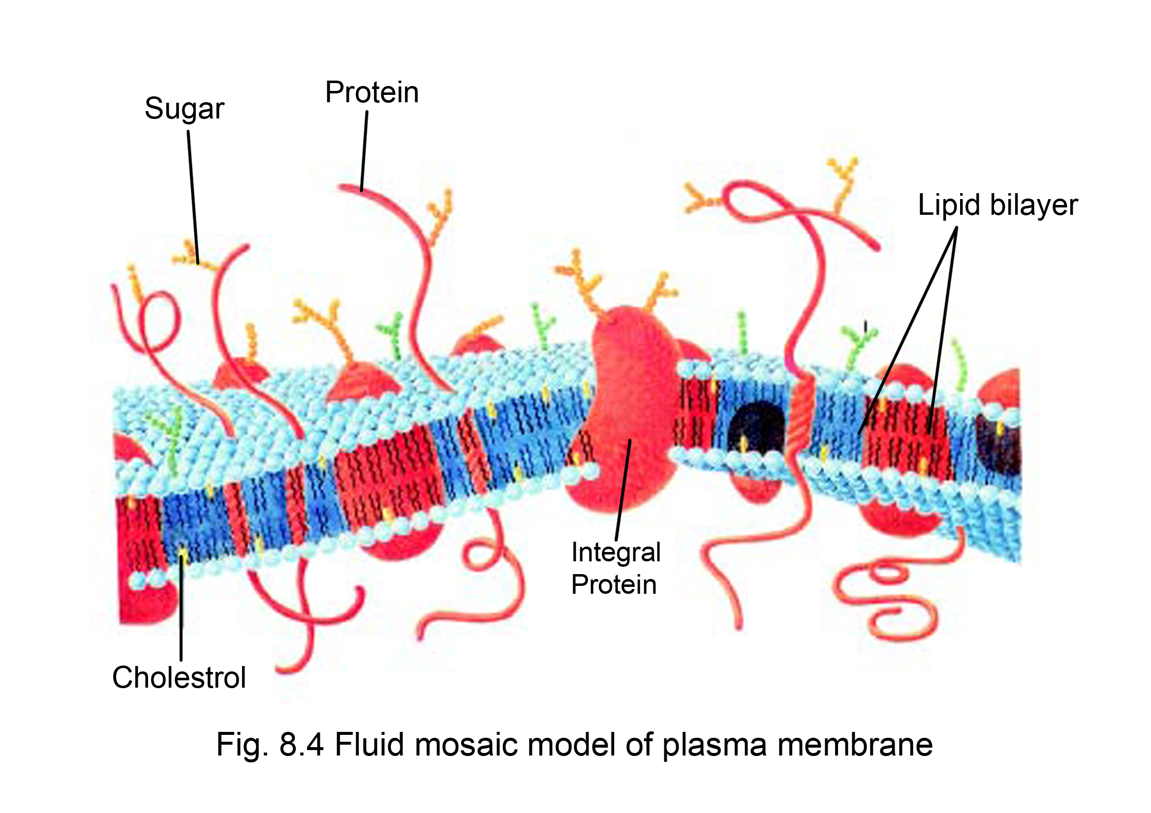
Some integral proteins span only part of the membrane. Single-pass integral proteins have a single membrane-spanning region consisting of 20–25 amino acids. Their hydrophobic membrane-spanning part interacts with the hydrophobic part of the bilayer. Integral proteins are entirely integrated into the membrane structure. Proteins are the second major component of the cell membrane and are randomly arranged within the lipid bilayer. It helps the cell membrane selectively permeable to larger molecules while allowing small molecules like carbon dioxide and oxygen to pass through easily. Cholesterol also prevents the compaction of the hydrophobic tails of lipids at low temperatures and the membrane’s expansion under heat. CholesterolĬholesterol is found within the phospholipids, allowing the membrane to retain its permeability and integrity even if there is a temperature change. This arrangement allows phospholipid molecules to form a bilayer structure that separates the cell’s interior from the exterior. Thus the surface of the cell membrane facing the interior and exterior of the cell is hydrophilic. Hydrophilic regions form hydrogen bonds with water and other polar molecules.

In an aqueous environment, the hydrophobic molecules arrange themselves to form a ball or a cluster. Each phospholipid molecule has a glycerol backbone with two fatty-acid molecules and a phosphate group attached to it. In contrast, the non-polar, hydrophobic tail is away from the aqueous environment. The polar, hydrophilic head is constantly in contact with the fluid inside and outside the cell. Phospholipids are amphiphilic, having water-attraction (hydrophilic) and water-repulsion (hydrophobic) regions. In this Special Issue, the use of membrane phospholipids to modify cellular membranes in order to modulate clinically relevant host properties is considered.Ĭytoskeletal interactions endosomes extracellular matrix lipid interactions lipid rafts membrane domains membrane dynamics membrane fusion membrane structure membrane vesicles.The model describes the structure of the cell membrane as a mosaic of these components having a thickness of 5-10 nm. They can also be externalized in a reverse process and released as extracellular vesicles and exosomes. Various lipid globules, droplets, vesicles and other membranes can fuse to incorporate new lipids or expel damaged lipids from membranes, or they can be internalized in endosomes that eventually fuse with other internal vesicles and membranes. However, the fluid regions of membranes are very important in lipid transport and exchange. The presence of specialized membrane domains significantly reduced the extent of the fluid lipid matrix, so membranes have become more mosaic with some fluid areas over time. In addition, lipid-lipid and lipid-protein membrane domains, essential for cellular signaling, were proposed and eventually discovered. Subsequently, the structures associated with membranes were considered, including peripheral membrane proteins, and cytoskeletal and extracellular matrix components that restricted lateral mobility. This simplified version of cell membrane structure was never proposed as the ultimate biomembrane description, but it provided a basic nanometer scale framework for membrane organization.


Integral membrane proteins can transform into globular structures that are intercalated to various degrees into a heterogeneous lipid bilayer matrix. The Fluid-Mosaic Membrane Model accounted for these and other properties, such as membrane asymmetry, variable lateral mobilities of membrane components and their associations with dynamic complexes. These thermodynamically untenable structures did not allow lipid lateral movements independent of membrane proteins. Early cell membrane models placed most proteins external to lipid bilayers in trimolecular structures or as modular lipoprotein units.


 0 kommentar(er)
